
Combination therapy for scalp angiosarcoma using bevacizumab and chemotherapy: a case report and review of literature
Introduction
Accounting for less than 2% of all soft tissue sarcomas, angiosarcoma (AS) is a rare malignant neoplasm derived from vascular endothelial cells (1-4). AS can occur in any region of the body; however, approximately 60% of tumors are cutaneous and are commonly found in the face and scalp region (2). AS is highly aggressive and has a poor prognosis. It can easily metastasize to local lymph nodes as well as distal organs due to its origin in the blood and lymphatic vessels. Specifically, lesions located on the scalp and face have a low survival rate and high recurrence rate (5), implying that AS at these sites may be more aggressive and refractory to therapy.
Due to the rapid progression of AS and the lack of a standard treatment modality, efficient management has long been a challenge. In general, combination chemotherapy using ifosfamide and doxorubicin has been used for the treatment of soft tissue sarcomas (2).
An increasing body of evidence has shown that bevacizumab (trade name: Avastin), a recombined humanized monoclonal antibody against vascular endothelial growth factor (VEGF), is a promising therapeutic option for AS management (6-8). It has been reported that bevacizumab, if used as a single agent, gives rise to a partial response in patients with extensive facial cutaneous AS (9). Application of bevacizumab in combination with radiation has shown promise in patients with AS of the scalp and head/neck (7,8). However, our understanding of the effectiveness of combined therapy of bevacizumab and chemotherapy agents is limited. To date, there has only been one study evaluating paclitaxel and bevacizumab in inoperable AS (7). The current report details a case of advanced scalp AS treated with bevacizumab in combination with the chemotherapy agents ifosfamide and epirubicin; paclitaxel and cisplatin.
Case report
A 59-year-old Chinese man was hospitalized because of a scalp lump for 5 months and intermittent cough and hemoptysis for 2 months. The purplish lump on the top of his head had been identified five months prior to hospitalization. At that time, the lump was approximately 1 cm in diameter and not painful, and no treatment was initiated. Three months after the lump was identified, the man presented with symptoms of intermittent cough, hemoptysis and bloody sputum. In addition, the lump was growing in size. Upon hospitalization, the physical examination revealed a purplish, intermediate hard lump of 2 cm × 3 cm, extending from the left side of the scalp to the upper forehead (Figure 1A). The lump on the left parotid gland was 2 cm × 2 cm in size. He had pain in his chest, abdomen, waist and legs, corresponding to a score of 6-7 using a visual analog scale (VAS). The tumor markers (assessed on November 2, 2010) were as follows: CEA, 0.46 ng/mL; CA-199, 4.20 U/mL; and CA-125, 11.9 U/mL. All of these were within the normal ranges. The histopathological report after scalp tumor excision showed that a piece of tissue 4.3 cm × 2.2 cm × 1.3 cm in size was excised, along with a piece of skin 4.3 cm × 2.2 cm in size. The diagnosis was angiosarcoma stage IV (Figure 1B), growing diffusely in the dermis and subcutaneous tissues. The immunohistochemical results were as follows: CD34 (+++), CD31 (+++), F8 factor (++), vimentin (+++), AAT (±), CK7 (–), HMB-45 (–), melan-A (–), melan-Pan (–), S-100 (–), CD68 (–), EMA (–), desmin (–), Bcl-2 (–), PR (–), and Ki-67 index (80%). A chest CT showed metastases to both lungs. PET-CT (October 25, 2010) showed tumor metastases to multiple organs, including the lungs, left parotid gland, mediastinum, hilar lymph nodes, liver, right peritoneum, and bones at multiple sites.
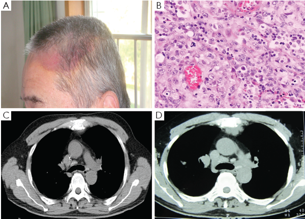
Combination therapy of bevacizumab and the chemotherapy drugs ifosfamide (IFO) and epirubicin (E-ADM) was started on November 5, 2010. The detailed treatment regimen was as follows: bevacizumab 5 mg/kg (300 mg) (i.v.) d1, repeated every 2 weeks; IFO 1.2 g/m2 (2 g) i.v. d1-4; mesna dose is 20% of the IFO dose (400 mg) i.v. t.i.d. d1-4; and epirubicin 30 mg/m2 i.v. d1-2. The cycle was repeated every 21 d. Physical examination after two cycles of treatment showed dramatic improvement in systemic pain and the tumor on the left parotid gland, as well as a reduction in scalp swelling. Additionally, a reduction in the size of the lung metastasis was observed by chest CT and scored as partial remission (PR). The original treatment regimen was continued for seven cycles. However, the chemotherapy was terminated due to bone marrow suppression, after which the patient only received bevacizumab 5 mg/kg (300 mg) every other week. After 2 months, the patient reported chest pain with a VAS score of 5-6. A follow-up chest CT revealed a grape-shaped soft tissue-like shadow behind the sternum and opacity of the bone boundary, indicating the presence of new lesions (Figure 1C,D). The treatment regimen was adjusted to the following: bevacizumab 5 mg/kg (300 mg) i.v. d1, repeated every 2 weeks; paclitaxel 150 mg/m2 (270 mg) i.v. d1; cisplatin 70 mg/m2 (120 mg) i.v. d2. The cycle was repeated every 21 d. The new therapy reduced the chest pain to a VAS score of 1-2. After receiving the new treatment for three cycles, the patient suddenly presented symptoms of chest tightness, suffocation, shortness of breath with cough, and difficulty expectorating. His heart rate increased to 140/min, while his blood oxygen decreased by 50-70% and he wheezed with each breath. Though immediate treatment was administered, the patient’s condition deteriorated sharply and he died of a pulmonary embolism.
Discussion
Soft tissue sarcomas contribute to less than 1% of all types of adult solid tumors and AS accounts for less than 1% of soft tissue sarcomas, making it a rare malignancy (4). While the traditional radio- and chemotherapies are commonly used, neo-adjuvant therapy for AS has started to attract a wide range of attention (10,11). The prognosis for early-stage AS corresponds to whether the bulk tumor has been completely surgically removed, and whether the tumor is larger than 5 cm. Studies report disease-free survival of 10-20% after 5 years (12-16) and less than 50% at 5 years (12-14,15-18). The approval of bevacizumab provides an additional therapeutic option for AS management (6-8).
AS is derived from vascular endothelial cells and has been reported to overexpress not only VEGF protein but also VEGF receptors (19-21). This implies that VEGF might be important for the development, progression, maintenance, and/or metastasis of this type of tumor. Therefore, it is logical to postulate that using bevacizumab to block the activity of VEGF will have an anti-growth effect on AS tumors, as it does on other solid tumors in colon, brain, kidney, and lung cancers (22). Indeed, clinical studies have shown that bevacizumab can significantly improve the survival of AS patients when used in combination with either radiation or the chemotherapy agent paclitaxel (7,8). However, our understanding of the effectiveness of combined therapy of bevacizumab and chemotherapy agents is still limited.
Given that the patient in this study had developed systemic metastasis prior to hospitalization, surgical cytoreduction did not seem practical. Therefore, the traditional chemotherapy agents ifosfamide and epirubicin (IFO + E-ADM) were used in combination with bevacizumab as the first-line treatment strategy. The addition of bevacizumab into the treatment regimen may have contributed to a short-term effect of partial remission and improved the patient’s quality of life. However, the patient developed bone marrow repression due to the infusion of IFO and E-ADM and the treatment was changed to paclitaxel, cisplatin and bevacizumab, which produced a similar response. Paclitaxel has been demonstrated to significantly enhance the disease-free survival in patients with head and neck or scalp AS (3) and is increasingly accepted as a first-line agent for AS.
The use of bevacizumab in conjunction with chemotherapy may improve quality of life and survival time in patients with metastasized AS. In this case of stage IV AS, the treatment reduced chest pain and resulted in 15 months of survival following the diagnosis. The patient died of acute pulmonary embolism (PE), one of the major forms of venous thromboembolism (VTE), which also includes deep vein thrombosis (DVT) (23). PE is the second leading cause of death in cancer patients in China (35%), which is significantly higher than the global rate 6% (23). Cancer is one of the main risk factors for VTE since tumor cells can secrete procoagulants that directly activate the coagulation system leading to thrombosis. Additionally, circulating tumor cells can become trapped within pulmonary capillaries resulting in initiation of the coagulation cascade and subsequent embolism. In particular, the vascular endothelial origin of AS makes it easier to disseminate tumor cells into the circulation and thereby cause embolism. Anti-cancer treatment, such as the administration of anti-angiogenic drugs like bevacizumab, could also contribute to VTE (24). The reported incidence of VTE in bevacizumab-treated cancer patients ranges from 3% to 23% (25). A recent meta-analysis reported an increased risk of VTE associated with bevacizumab therapy among cancer patients [relative risk 1.33 (95% confidence interval 1.13-1.56); P<0.001] (26). However, whether or not the use of bevacizumab contributed to PE in this case is unknown. The benefits of bevacizumab treatment for AS will need to be weighed against the risks of PE in this population.
Acknowledgements
This work is supported by the Wu Jieping Medical Foundation Grant (No. 320.6750.11074).
Disclosure: The authors declare no conflict of interest.
References
- Lydiatt WM, Shaha AR, Shah JP. Angiosarcoma of the head and neck. Am J Surg 1994;168:451-4. [PubMed]
- Aust MR, Olsen KD, Lewis JE, et al. Angiosarcomas of the head and neck: clinical and pathologic characteristics. Ann Otol Rhinol Laryngol 1997;106:943-51. [PubMed]
- Fury MG, Antonescu CR, Van Zee KJ, et al. A 14-year retrospective review of angiosarcoma: clinical characteristics, prognostic factors, and treatment outcomes with surgery and chemotherapy. Cancer J 2005;11:241-7. [PubMed]
- Young RJ, Brown NJ, Reed MW, et al. Angiosarcoma. Lancet Oncol 2010;11:983-91. [PubMed]
- Maddox JC, Evans HL. Angiosarcoma of skin and soft tissue: a study of forty-four cases. Cancer 1981;48:1907-21. [PubMed]
- Koontz BF, Miles EF, Rubio MA, et al. Preoperative radiotherapy and bevacizumab for angiosarcoma of the head and neck: two case studies. Head Neck 2008;30:262-6. [PubMed]
- Fuller CK, Charlson JA, Dankle SK, et al. Dramatic improvement of inoperable angiosarcoma with combination paclitaxel and bevacizumab chemotherapy. J Am Acad Dermatol 2010;63:e83-4. [PubMed]
- De Yao JT, Sun D, Powell AT, et al. Scalp Angiosarcoma Remission with Bevacizumab and Radiotherapy without Surgery: A Case Report and Review of the Literature. Sarcoma 2011;2011:160369.
- Rosen A, Thimon S, Ternant D, et al. Partial response to bevacizumab of an extensive cutaneous angiosarcoma of the face. Br J Dermatol 2010;163:225-7. [PubMed]
- Espat NJ, Lewis JJ, Woodruff JM, et al. Confirmed angiosarcoma: prognostic factors and outcome in 50 prospectively followed patients. Sarcoma 2000;4:173-7. [PubMed]
- Park MS, Ravi V, Araujo DM. Inhibiting the VEGF-VEGFR pathway in angiosarcoma, epithelioid hemangioendothelioma, and hemangiopericytoma/solitary fibrous tumor. Curr Opin Oncol 2010;22:351-5. [PubMed]
- Hodgkinson DJ, Soule EH, Woods JE. Cutaneous angiosarcoma of the head and neck. Cancer 1979;44:1106-13. [PubMed]
- Holden CA, Spittle MF, Jones EW. Angiosarcoma of the face and scalp, prognosis and treatment. Cancer 1987;59:1046-57. [PubMed]
- Freedman AM, Reiman HM, Woods JE. Soft-tissue sarcomas of the head and neck. Am J Surg 1989;158:367-72. [PubMed]
- Willers H, Hug EB, Spiro IJ, et al. Adult soft tissue sarcomas of the head and neck treated by radiation and surgery or radiation alone: patterns of failure and prognostic factors. Int J Radiat Oncol Biol Phys 1995;33:585-93. [PubMed]
- Mark RJ, Poen JC, Tran LM, et al. Angiosarcoma. A report of 67 patients and a review of the literature. Cancer 1996;77:2400-6. [PubMed]
- Farhood AI, Hajdu SI, Shiu MH, et al. Soft tissue sarcomas of the head and neck in adults. Am J Surg 1990;160:365-9. [PubMed]
- Ferrara N, Gerber HP, LeCouter J. The biology of VEGF and its receptors. Nat Med 2003;9:669-76. [PubMed]
- Stacher E, Gruber-Mösenbacher U, Halbwedl I, et al. The VEGF-system in primary pulmonary angiosarcomas and haemangioendotheliomas: new potential therapeutic targets? Lung Cancer 2009;65:49-55. [PubMed]
- Tokuyama W, Mikami T, Masuzawa M, et al. Autocrine and paracrine roles of VEGF/VEGFR-2 and VEGF-C/VEGFR-3 signaling in angiosarcomas of the scalp and face. Hum Pathol 2010;41:407-14. [PubMed]
- Liu W, Xu J, Wang M, et al. Tumor-derived vascular endothelial growth factor (VEGF)-a facilitates tumor metastasis through the VEGF-VEGFR1 signaling pathway. Int J Oncol 2011;39:1213-20. [PubMed]
- Reck M, von Pawel J, Zatloukal P, et al. Overall survival with cisplatin-gemcitabine and bevacizumab or placebo as first-line therapy for nonsquamous non-small-cell lung cancer: results from a randomised phase III trial (AVAiL). Ann Oncol 2010;21:1804-9. [PubMed]
- Jenkins EO, Schiff D, Mackman N, et al. Venous thromboembolism in malignant gliomas. J Thromb Haemost 2010;8:221-7. [PubMed]
- Norden AD, Bartolomeo J, Tanaka S, et al. Safety of concurrent bevacizumab therapy and anticoagulation in glioma patients. J Neurooncol 2012;106:121-5. [PubMed]
- Zangari M, Fink LM, Elice F, et al. Thrombotic events in patients with cancer receiving antiangiogenesis agents. J Clin Oncol 2009;27:4865-73. [PubMed]
- Nalluri SR, Chu D, Keresztes R, et al. Risk of venous thromboembolism with the angiogenesis inhibitor bevacizumab in cancer patients: a meta-analysis. JAMA 2008;300:2277-85. [PubMed]